cameron ulcer causes
Though typically asymptomatic these may rarely present as acute severe upper gastrointestinal bleed GIB. Later this type of ulcer was named after his name Cameron lesions.
Peptic Ulcers Smartengage
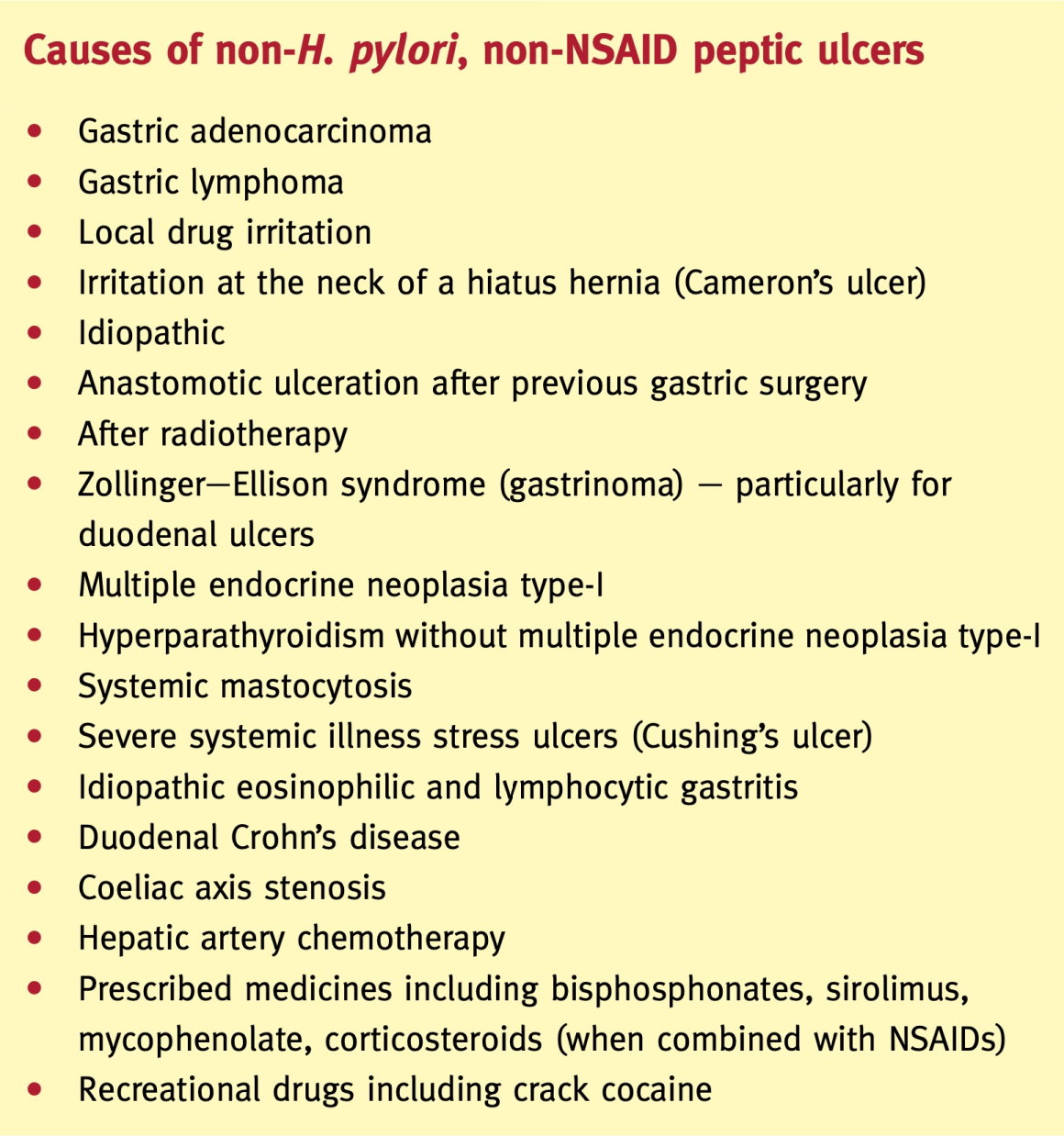
An infection with the bacteria Helicobacter pylori.

. Concomitant acid-peptic diseases are seen in a majority of individuals especially reflux esophagitis and its complications. Cameron lesions are linear gastric ulcers or erosions on the mucosal folds at the diaphragmatic impression in patients with a large hiatal hernia. Cameron suggested mechanical trauma as the cause because the gastric folds at the level of constriction of the diaphragm rubbed against each other.
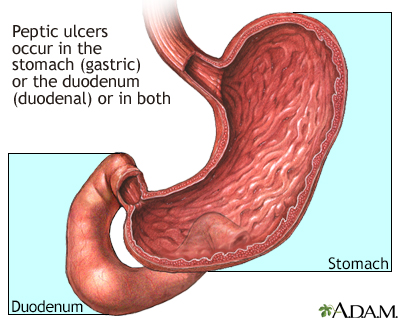
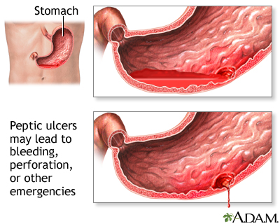
Causes of peptic ulcers include. Such lesions may be found in upto 50 of endoscopies performed for another indication. The most common causes of upper GI bleed include peptic ulcer disease gastroesophageal varices esophagitis angioectasia and vascular lesions.
The choice of therapy of Cameron lesions medical or surgical should be individualized for each patient. The most common causes of upper GI bleed include peptic ulcer disease gastroesophageal varices esophagitis angioectasia and vascular lesions. The diagnosis is usually made during upper endoscopy.
Long-term use of nonsteroidal anti-inflammatory drugs such as aspirin and ibuprofen. The etiology is unknown in about 8 of the cases. In 1986 Dr.
7 Based on observations of the stomach in 450 patients undergoing open surgery for hernia repair Colin et al 5 suggested that the pressure difference between the abdomen and thorax caused a sliding movement of. Cameron lesions are erosions or ulcerations commonly found in patients with hiatal hernias which can lead to both chronic and at times life-threatening acute bleeding. The most common causes.
Cameron lesions are linear gastric ulcers or erosions on the mucosal folds at the diaphragmatic impression in patients with a large hiatal hernia. Some propose that Cameron lesions form as a consequence of local mechanical trauma from gastric folds rubbing against each other as the hernia slides up and down past the diaphragmatic hiatus whereas others propose that these erosions could be related to Helicobacter pylori infection transient ischemia acid reflux and gastric or vascular stasis. Cameron lesions have been reported at a prevalence rate between 33 and 52 in patients with hiatal hernias undergoing esophagogastroduodenoscopy EGD however those without evidence of hiatal.
Iatrogenic bleeding after endoscopic interventions. These ulcers occur when a person has a large hiatal hernia and often cause GI bleeding. Some propose that Cameron lesions form as a consequence of local mechanical trauma from gastric folds rubbing against each other as the hernia slides up and down past the diaphragmatic hiatus whereas others propose that these erosions could be related to Helicobacter pylori infection transient ischemia acid reflux and gastric or vascular stasis.
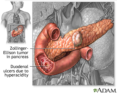
Peptic ulcer disease is a common cause of gastrointestinal bleeding and is usually related to Helicobacter pylori H. Rare cancerous and noncancerous tumors in the stomach duodenum or pancreas known as Zollinger-Ellison syndrome. An often overlooked cause of iron deficiency anaemia in patients with large hiatal hernias.
Cameron lesions represent linear gastric erosions and ulcers on the crests of mucosal folds in the distal neck of a hiatal hernia HH. Of all the GI hemorrhages nearly 50 are due to upper GI bleeding. The clinical relevance of Cameron lesions is due to their potential complications such as gastrointestinal bleeding acute chronic and obscure and anemia.
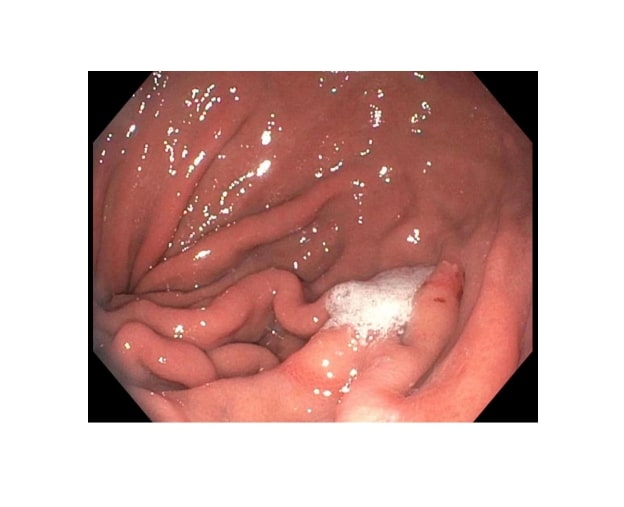
Cameron lesion is a rare cause of occult upper GI bleed. A Cameron lesion is a linear erosion or ulceration of the mucosal folds lining the stomach where it is constricted by the thoracic diaphragm in persons with large hiatal hernias. Gastric ischemia and stasis.
Mechanical trauma ischemia and acid mucosal injury may play a role in the pathogenesis of Cameron lesions. Another rare type of ulcer is called Camerons ulcer. Cameron lesions are non-peptic non-gastroesophageal reflux disease-associated mucosal defects which develop on the top of gastric.
Pylori infection or nonsteroidal anti-inflammatory drugs. Gastric antral vascular ectasia. Treatment of anemia with Cameron lesions includes iron supplements and acid.
Cameron lesion is a rare cause of occult upper GI bleed. The lesions may cause chronic blood loss resulting in iron deficiency anemia. Showing results for cameron ulcer Causes of upper gastrointestinal bleeding in adultslesion.
Less often they cause acute bleeding. Though typically asymptomatic several complications can occur including gastroesophageal reflux disease 3 iron deficiency anemia 4 acute or chronic bleeding 5 and ulcer or erosion formation 2. Cameron lesions can cause weird symptoms with a Hiatal hernia such as fatigue dizziness fast heartbeats and shortness of breath.
Cameron first described linear elongated stomach ulcers associated with large Hiatal hernias. The lesions are associated with occult bleeding and development of chronic iron deficiency anaemia but are. The etiology is unknown in about 8 of the cases.
Usually an incidental endoscopic finding Cameron lesions represent linear gastric erosions and ulcers on the crests of mucosal folds in the distal neck of. Patients with a large hiatal hernia are at risk for a Cameron ulcer which.

Helicobacter Pylori Infection And Peptic Ulcers Medicine

Histopathological Characterization Of A Cameron Lesion Semantic Scholar
Cameron Lesions Article

Gastric Ulcers Iv The Gastrointestinalatlas Gastrointestinalatlas Com
Causes Of Peptic Ulcer Disease H Pylori Nsaids Grepmed

Cameron Lesions And Disorders Of The Cardia Human Anatomy Nuclear Imaging Digestive System
2

2021 European Guideline For The Management Of Vulval Conditions Meijden 2022 Journal Of The European Academy Of Dermatology And Venereology Wiley Online Library

Histopathological Characterization Of A Cameron Lesion Semantic Scholar

Cameron Lesions Wikiwand

Cameron Ulcer Causing Severe Anemia In A Patient With Diaphragmatic Hernia Abstract Europe Pmc
In Depth Reports Peptic Ulcers

Cirbosque Some4surgery Twitter પર Pictorial Explanation Of The Cameron Ulcers In A Paraesophageal Hernia By Dr Frank Netter Some4surgery Ascolcx2020 Medtwitter Juliomayol Swexner Pferrada1 Pipecabrerav Almagoch Drthawaba Neilflochmd
Pdf An Unusual Cause Of Anemia Cameron Ulcer
Peptic Ulcers Smartengage

A Life Threatening Problem Hidden Within A Hiatal Hernia Medpage Today

Peptic Ulcer Disease Mallappa Shalavadi

Peptic Ulcer Disease

Histopathological Characterization Of A Cameron Lesion Semantic Scholar


Comments
Post a Comment